IMAGE ATLAS

About the Site:

The thyroid web atlas contains 300 images of key thyroid lesions in an easy-to-search format. All images have been independently reviewed and approved by the committee members.

Using the Atlas:

  • Click on an album to see the thumbnail images
  • Click on the thumbnails to see the intermediate images
  • Click on the intermediate image to obtain the full sized image

Use of Images:

These images are intended for educational purposes and may be freely used for such as long as the Papanicolaou Society and the Bethesda System for Reporting Thyroid Cytopathology is credited.

  Gallery Links - Home :: Login :: Album list :: Last uploads :: Last comments :: Most viewed :: Top rated :: My Favorites :: Search
   
PSCO Website Links - Atlas Home - Main - About Us - News - Case of the Month - Guidelines - Links - Contact
Home > FOLLICULAR NEOPLASM or SUSPICIOUS FOR A FOLLICULAR NEOPLASM

Most viewed - FOLLICULAR NEOPLASM or SUSPICIOUS FOR A FOLLICULAR NEOPLASM
follicular_neoplasm_hurthle_cell_type-fu_hurthle_cell_carcinoma-pap19e-high-sturgis.jpg
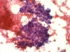
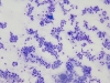
Crowded follicular cells.40644 views
follicular_neoplasm_hurthle_cell_type-fu_hurthle_cell_carcinoma-pap15-high-sturgis.jpg
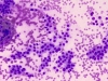
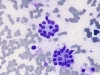
Crowded follicular cells and microfollicle.18192 views
follicular_neoplasm_hurthle_cell_type-fu_hurthle_cell_carcinoma-pap19b-high-sturgis.jpg
Follicular cells in trabecular arrangement.17604 views
image_000302_004~1.JPG
Capillaries.15340 views
follicular_neoplasm_hurthle_cell_type-fu_hurthle_cell_carcinoma-pap14-high-sturgis.jpg
Marked follicular cell crowding.15021 views
image_000305_001~1.JPG
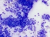
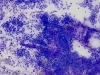
Very cellular specimen with trabecular arrangements (ribbons) of follicular cells and minimal colloid.14910 views
image_000302_003~1.JPG
Very cellular specimen with numerous microfollicles and minimal colloid.13156 views
image_000305_002~1.JPG
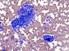
Crowded follicular cells and minimal colloid.12865 views
image_000302_001~1.JPG
Very cellular specimen with numerous microfollicles and minimal colloid.12413 views
image_000305_003~1.JPG
Microfollicles.12187 views
image_000302_002~1.JPG
Very cellular specimen with numerous microfollicles and minimal colloid.12168 views
follicular_neoplasm_hurthle_cell_type-fu_hurthle_cell_adenoma-dq10-high-sturgis.jpg
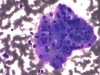
Cohesive fragment of Hurthle cells (oncocytes).9947 views
30 files on 3 page(s) 1