IMAGE ATLAS

About the Site:

The thyroid web atlas contains 300 images of key thyroid lesions in an easy-to-search format. All images have been independently reviewed and approved by the committee members.

Using the Atlas:

  • Click on an album to see the thumbnail images
  • Click on the thumbnails to see the intermediate images
  • Click on the intermediate image to obtain the full sized image

Use of Images:

These images are intended for educational purposes and may be freely used for such as long as the Papanicolaou Society and the Bethesda System for Reporting Thyroid Cytopathology is credited.

  Gallery Links - Home :: Login :: Album list :: Last uploads :: Last comments :: Most viewed :: Top rated :: My Favorites :: Search
   
PSCO Website Links - Atlas Home - Main - About Us - News - Case of the Month - Guidelines - Links - Contact
Most viewed
Ed82C_1~0.jpg
Nuclear crowding and powdery chromatin.4475 views
C01-23782D.jpg
Finely textured chromatin and intranuclear pseudoinclusions.4472 views
BC06-44681D.jpg
Some tumor cells appear glandular, with granular cytoplasm, round nuclei, and prominent nucleoli (ThinPrep).4424 views
Cystic_PTC-5.jpg
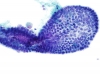
Papillary Carcinoma of Thyroid - Cystic Variant4410 views
BS07-16211E.jpg
Histologic section.4406 views
Flame.jpg
Flame Cells in Grave's Disease4401 views
ED62A.jpg
Intranuclear pseudoinclusions and focal oncocytic change.4399 views
BC06-44681E.jpg
Tumor cells exhibit squamous differentiation with keratin formation (ThinPrep).4397 views
C00-4582B.jpg
Intact papilla.4381 views
BC06-44681C.jpg
Intermediate-type cells and small keratin pearls (ThinPrep).4346 views
Papillary_CA,_giant_cell,_DQ_lp.jpg
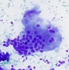
Multinucleated giant cell in papillary carcinoma4341 views
C01-23782C.jpg
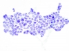
Monolayered sheet.4338 views
334 files on 28 page(s) 21